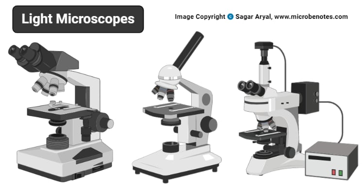
Light Microscope Source Of Radiation
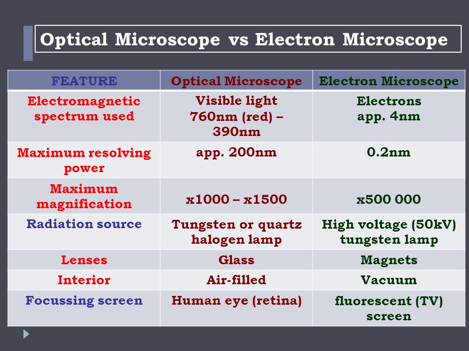
Differences Between Light Microscope And Electron Microscope

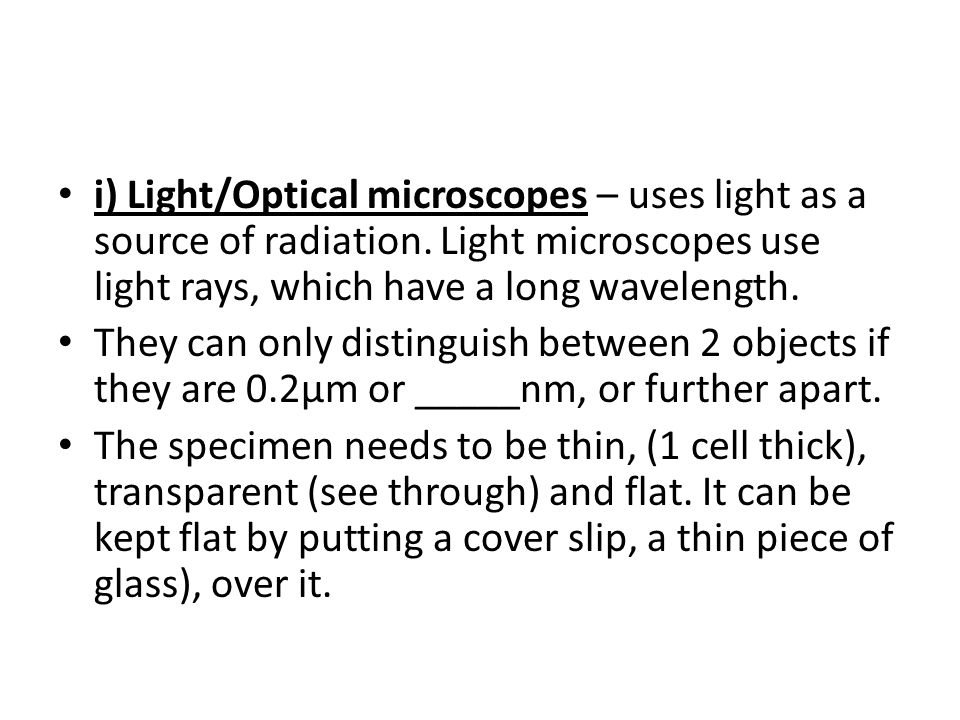
Light Microscopes An Overview Sciencedirect Topics
Histological Techniques 6 Light Microscope Atlas Of Plant And Animal Histology
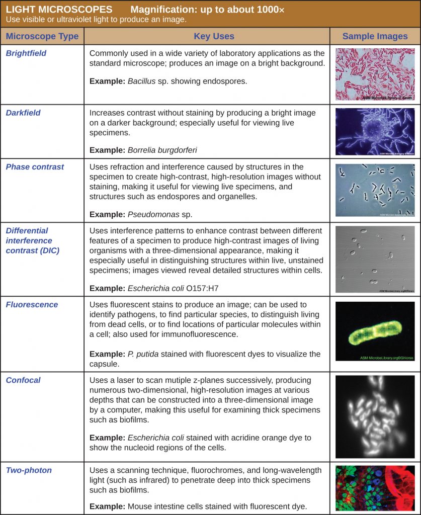
Instruments Of Microscopy Microbiology
2 3 Instruments Of Microscopy Biology Libretexts
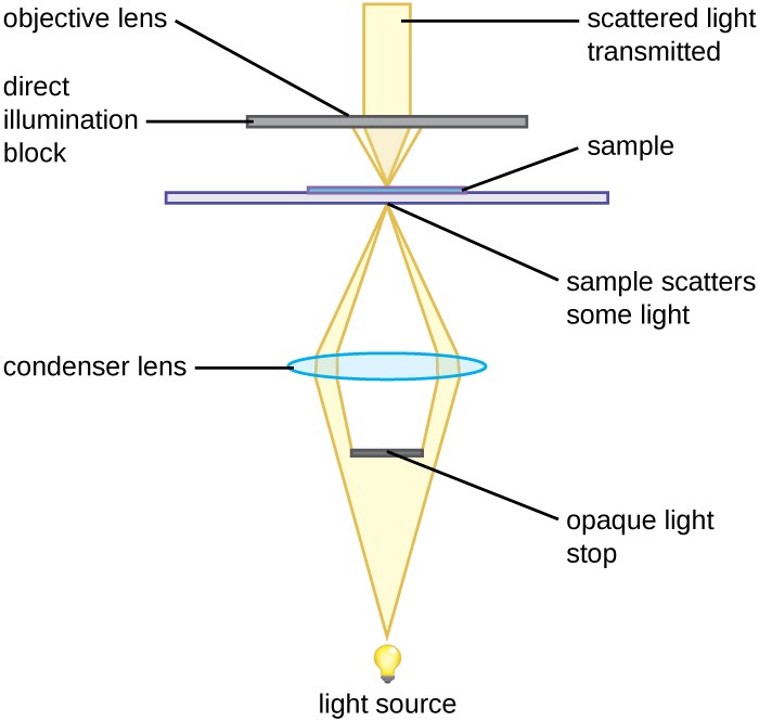
Instruments Of Microscopy Microbiology
In general achromat lenses are the most basic whereas plan apochromats are often considered superior.
Light microscope source of radiation.
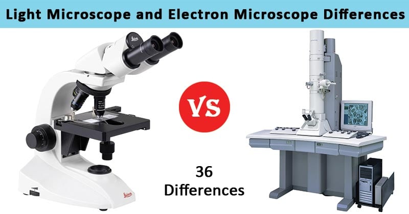
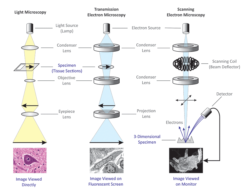
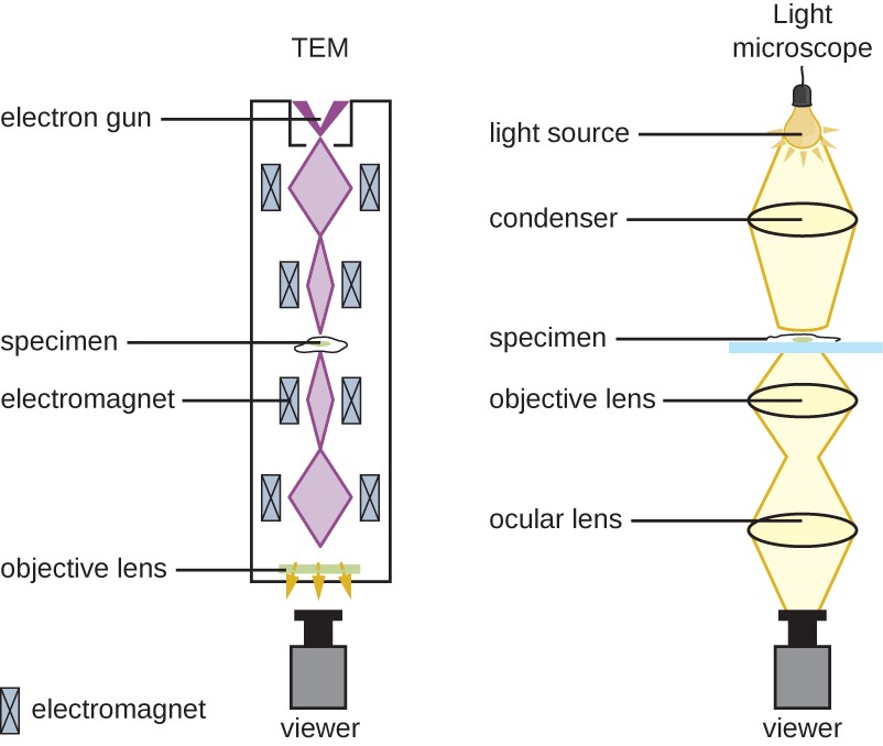
36 Differences Between Light And Electron Microscope

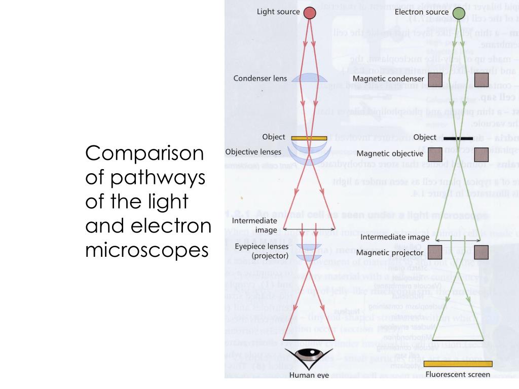
Contrast The Way Light Microscopes And Electron Microscopes Magnify Objects
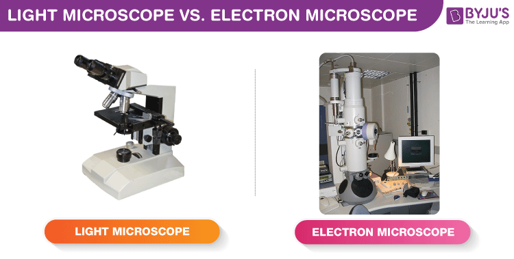
Difference Between Light Microscope And Electron Microscope Byju S

The Electron Microscope Introduction Ppt Download
Light Microscopy Central Microscopy Research Facility
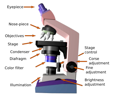
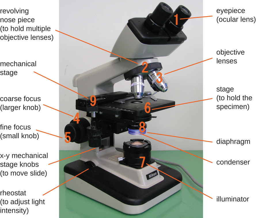
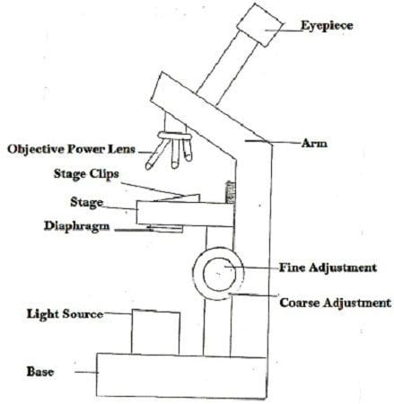
Light Microscope Definition Principle Types Parts Magnification
Cells And Microscopy What Is Magnification And Resolution Ppt Download
Difference Between Light Microscope And Electron Microscope With Advantages Disadvantages Its Types And Comparision Chart Bio Differences
Microscopic Observations Of Microorganisms Muhammed Mahfuzur Rahman Lecturer Department Of Pharmacy Ppt Download
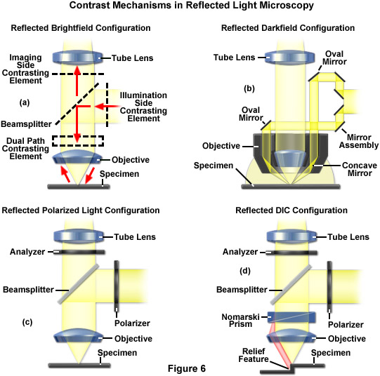
Zeiss Microscopy Online Campus Microscopy Basics Reflected Light Microscopy

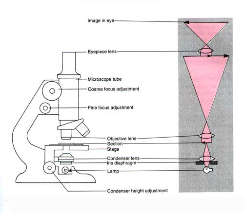
Principles Of Light Microscopy With A Compound Light Microscope We Can Examine Very Small Specimens As Well As Some Of Their Fine Detail A Series Of Ppt Download

Microscopy
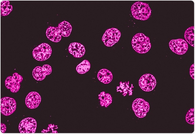
Fluorescence Microscopy Vs Light Microscopy
2 3 Instruments Of Microscopy Microbiology Canadian Edition
Transmission Electron Microscopy Central Microscopy Research Facility
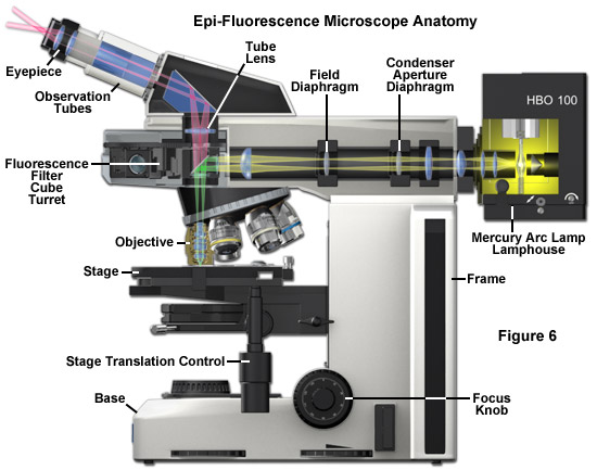
Zeiss Microscopy Online Campus Microscopy Basics Fluorescence Microscopy
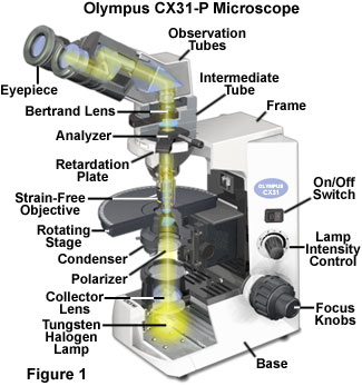
Polarized Light Microscopy Cx31 P Polarized Light Microscope Configuration Olympus Life Science
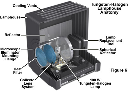
Zeiss Microscopy Online Campus Tungsten Halogen Lamps
Https Encrypted Tbn0 Gstatic Com Images Q Tbn 3aand9gctbgbjzegiwhzbcfxndgqughgbqunqiemqf Dbbdkuzginnodys Usqp Cau

Cells Origins

Optical Microscope An Overview Sciencedirect Topics
What Is Histology The Histology Guide

Compare Light Microscopes With Electron Microscopes As Biology Electron Microscope Scanning Electron Microscope Cell Organelles

Image Result For Compound Light Microscope Parts Microscope Parts Microscopic Body Tube

Electron Microscopes An Overview Sciencedirect Topics

What Are The Main Differences Between An Sem An Esem An Sem Fib And An S Tem Horiba
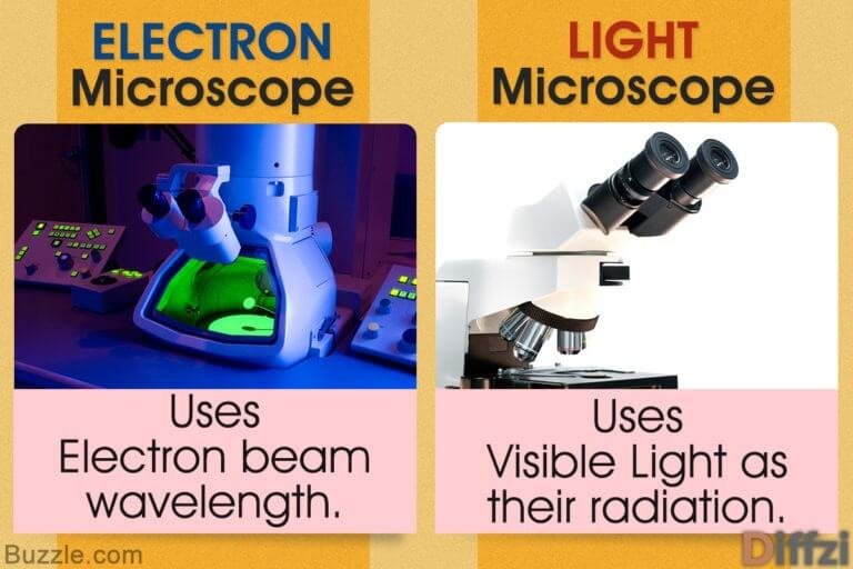
Light Microscope Vs Electron Microscope What Is The Difference Diffzi

Electron Microscopes Siyavula Textbooks Grade 12 Physical Science Openstax Cnx

Light Field Microscopy Wikipedia

Molecular Expressions Microscopy Primer Anatomy Of The Microscope Light Sources For Optical Microscopy

What Is Confocal Laser Scanning Microscopy

Chapter 2 Flashcards Quizlet

Cell Structure A Biology

Transmission Electron Microscope Tem Introduction To Jeol Products Jeol Ltd

On Line Biology Resources Use Of Dissection Microscopes

Tutorial Peem Technique
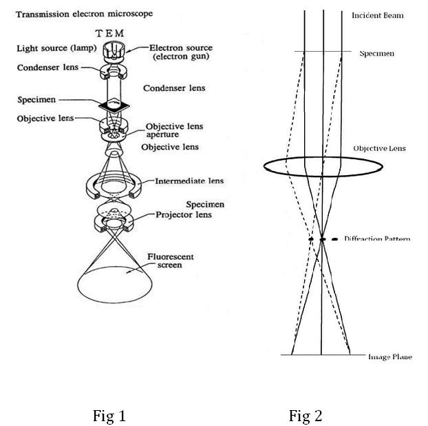
Transmission Electron Microscopy Tem
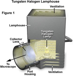
Kohler Illumination Light Sources Olympus Life Science

Electron Microscopes Vs Optical Light Microscopes Microbehunter Microscopy
Ppt Cell Structure Powerpoint Presentation Free Download Id 5516764
Uv Light Microscope Proves Useful Diagnostic Tool For Pathologists

Interference Microscopy An Overview Sciencedirect Topics

Biol2060 Cell Biology
1
Source : pinterest.com
